哺乳動物生物發(fā)光,一般是將Firefly luciferase基因(由554個氨基酸構成,約50KD)即熒光素酶基因整合到預期觀察的細胞染色體DNA上以表達熒光素酶,培養(yǎng)出能穩(wěn)定表達熒光素酶的細胞株,當細胞分裂、轉移、分化時, 熒光素酶也會得到持續(xù)穩(wěn)定的表達。基因、細胞和活體動物都可被熒光素酶基因標記。將標記好的細胞接種到實驗動物體內后,當外源(腹腔或靜脈注射)給予其底物熒光素(luciferin),即可在幾分鐘內產生發(fā)光現象。這種酶在ATP,氧存在的條件下,催化熒光素的氧化反應才可以發(fā)光,因此只有在活細胞內才會產生發(fā)光現象,并且發(fā)光光強度與標記細胞的數目線性相關。
熒光素由于諸多優(yōu)點得到廣大科研人員的青睞,主要特點如下:
(1) 熒光素不會影響動物的正常生理功能。
(2)熒光素是280道爾頓的小分子,水溶性和脂溶性都非常好,很容易穿透細胞膜和血腦屏障。
(3) 熒光素在體內擴散速度快,可通過腹腔注射或尾部靜脈注射進入動物體內。腹腔注射擴散較慢,持續(xù)發(fā)光長。熒光素腹腔注射老鼠后約1min后表達熒光素酶的細胞開始發(fā)光,10min后強度達到穩(wěn)定的最高點,在最高點持續(xù)約20~30 min后開始衰減,約3h后熒光素排除,發(fā)光全部消失,最佳檢測時間是在注射后15~35min之間;若進行熒光素靜脈注射,擴散快,但發(fā)光持續(xù)時間很短。科研人員根據大量的實驗總結出熒光素的合適的用量是150mg/kg,即體重20克的小鼠需要3毫克的熒光素。
(4) 觀察時間的間隔沒有最短限制,只要觀察的條件控制一致就可以。雖然底物在動物體內有一定的代謝過程,但是上一次底物的殘留曲線可以知道,可以控制對下一次觀察結果的影響。
光學原理:光在哺乳動物組織內傳播時會被散射和吸收,光子遇到細胞膜和細胞質時會發(fā)生折射現象,而且不同類型的細胞和組織吸收光子的特性并不一樣。血紅蛋白(hemoglobin)是造成體內可見光被吸收的主要因素,其吸收可見光中藍綠光波段的大部分。但是在可見光大于600納米的紅光波段,血紅蛋白的吸收作用卻很小。因此,在偏紅光區(qū)域, 大量的光可以穿過組織和皮膚而被檢測到。利用活體動物生物發(fā)光成像技術最少可以檢測到皮下的幾百個細胞。當然,由于發(fā)光源在老鼠體內深度的不同可看到的最少細胞數是不同的。一般認為,每一厘米深度,發(fā)光強度衰減10倍,血液豐富的組織或器官(比如心臟、肝臟、肺臟)衰減多,與骨骼相鄰的組織或器官衰減少。在相同的深度情況下,檢測到的發(fā)光強度和細胞的數量具有非常好的線性關系,可由儀器量化檢測到的光強度,反映出細胞的數量。
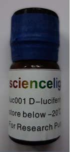
熒光素酶的發(fā)光是生物發(fā)光,不需要激發(fā)光,但需要底物熒光素(D-Luciferin,鉀鹽)。熒光素酶有554個氨基酸,約50KD。熒光素酶的底物熒光素,約280道爾頓。熒光素的水溶性和脂溶性都非常好,很容易穿透細胞膜和血腦屏障。熒光素是腹腔注射或尾部靜脈注射進入小鼠體內的,約一分鐘就可以擴散到小鼠全身。大部分發(fā)表的文章中,熒光素的濃度是150mg/kg 。20克的小鼠需要3毫克的熒光素。常用方法是腹腔注射,這種方法擴散較慢、開始發(fā)光慢、持續(xù)發(fā)光長。若進行熒光素靜脈注射,這種方法擴散快、開始發(fā)光快,但發(fā)光持續(xù)時間很短。熒光素(luciferin)在氧氣、ATP存在的條件下和熒光素酶發(fā)生反應,生成氧化熒光素(oxyluciferin),并產生發(fā)光現象。熒光素不會影響動物的正常生理功能。我們提供1g,400mg以及200mg 的包裝,價格優(yōu)惠。
常用的有兩種熒光素酶,Luciferase 和 Renilla熒光素酶,二者的底物不一樣,前者的底物是D-luciferin,后者的底物是coelentarizine (我們也有,如果需要請聯系)。二者的發(fā)光顏色不一樣,前者所發(fā)的光波長在540-600nm,后者所發(fā)的光波長在460-540nm左右。前者所發(fā)的光更容易透過組織,后者在體內的代謝比前者快。大部分的發(fā)表文章通常使用前者用作報告基因, 也有一些文章使用兩者進行雙標記。
熒光素腹腔注射老鼠后約一分鐘后表達熒光素酶的細胞開始發(fā)光,十分鐘后強度達到穩(wěn)定的最高點。在最高點持續(xù)約20-30 分鐘后開始衰減,約三小時后熒光素排除,發(fā)光全部消失。最好的檢測時間是在注射后15到35分鐘之間。
熒光素酶基因是插到細胞染色體內的,當細胞分裂、轉移、分化時, 熒光酶也會得到持續(xù)穩(wěn)定的表達。熒光素酶的半衰期約三個小時, 所以只有活細胞才能夠持續(xù)表達熒光素酶。
實驗方法如下:
Protocol 1: In Vitro Imaging of MDA-MB-231-luc and HCT116-Luc cells
Materials needed:
MDA-MB-231-luc cells (1 T175 flask)
HCT116-Luc (1 T75 flask)
Trypsin/EDTA
D-Luciferin Firefly, potassium salt(Sciencelight, Catalog# luc001), 1.0 g /vial,
Sterile water
Complete media for each cell line
96 well black plates (Costar)
Procedure:
A. Luciferin Preparation
1. Prepare a 200X luciferin stock solution (30 mg/ml) in sterile water. Mix gently by inversion until luciferin is completely dissolved. Use immediately, or aliquot and freeze at -20C for future use.
* Note: Reconstitute the quantity of D-Luciferin necessary for an individual experiment.
2. Prepare 2X luciferin (300ug/ml) in complete media. Quick thaw 200X stock solution and dilute 1:100 in complete media (2X solution)
* Note: This working solution should be prepared fresh for each experiment.
B. Cell Preparation and Serial Dilution
1. Trypsinize a flask and resuspend cells in a total of 10ml media.
2. Count cells and adjust cell concentration to 100,000 cells/ml = 10,000 cells/100ul.
3. Perform a serial dilution (1:2) in complete media as follows:
a. Pipette 200 ul cells into well #1 (in duplicate).
b. Add 100 ul media to wells #2-10 (in duplicate) using a multi-channel pipette.
c. Perform 1:2 serial dilution by pipetting 100ul from well #1 to well #2 using a multi-channel pipette, mixing well, then taking 100 ul from well 2 to well 3. Continue dilution to well #10 and discard 100 ul from well #10 after mixing.
C. Imaging Procedure
1. Add 100 ul 2X luciferin to cells in well #1-10.
2. Place plate of cells in the camera system on FOV15cm to view entire plate. Use the Plate Positioner or Alignment Grid to help position the plate.
3. Approximately 2-3 minutes after the addition of luciferin, image plate at 1 minute, 10 bin.
* Note: If using >10,000 cells in well #1, imaging time should de decreased.
D. Data Analysis
1. Apply grid ROI to image; measure photons/well.
2. Export measurements from LI lab book to an excel file.
3. Are dilutions consistent? What is the minimum number of cells detected?
Protocol 2: In Vivo imaging of MDA-MB-231-luc and HCT116-Luc Subcutaneous Model
Materials:
MDA-MB-231-Luc and HCT116-Luc cells
Mice: 2 subcutaneous HCT116-Luc tumor-bearing female BALB/c nude mice;
4 naïve female BALB/c nude mice;
Artagain black paper (Strathmore, Catalog# 445-109)
D-Luciferin Firefly, potassium salt(Sciencelight, Catalog# luc001), for in vivo imaging
Syringe (1ml) and needles (25 x 5/8" gauge)
Anesthesia (Pentobarbital Sodium)
Procedure:
A. Luciferin Preparation
1. Prepare a stock solution of luciferin at 15mg/ml in DPBS. Filter sterilize through a 0.2 um filter.
2. Prepare enough to inject 10 ul/g of body weight. Each mouse should receive 150 mg luciferin/kg body weight. (e.g. For a 20 g mouse, inject 200 ul to deliver 3 mg of luciferin.)
B. Preparation of tumor cells:
1. Trypsinize the cells from flasks, wash once in serum-free medium.
2. Resuspend in room temperate serum-free medium to a concentration of 1x10⁶ cells in 100ul. Hold at room temperature until ready for use.
C. Injections:
1. Inject mice with firefly luciferin (150 mg/kg) by intraperitoneal injection using a 25 x 5/8” gauge needle.
2. After 7-8 minutes, anesthetize mice by intraperitoneal injection of pentobarbital sodium using a 25 x 5/8” gauge needle.
3. (1) Inject tumor cells (1x10⁶ cells in 100ul) subcutaneously into the dorsal flank of the mouse.
(2) For the two HCT116-Luc tumor-bearing mice, no cells were inoculation to the mice.
D. Imaging
Place mice on black paper in the imaging box and image dorsally to detect tumor cells.
Protocol 3: In Vivo imaging of HCT116-Luc Intravenous Model
Materials:
HCT116-Luc cells
Mice: 1 naïve female BALB/c nude mice
Artagain black paper (Strathmore, Catalog# 445-109)
D-Luciferin Firefly, potassium salt(Sciencelight, Catalog# luc001), for in vivo imaging
Syringe (1ml) and needles (25 x 5/8" gauge and 26 x 1/2" gauge)
Heating lamp and mouse restrainer (for cell injection)
Anesthesia (Pentobarbital Sodium)
Procedure:
A. Luciferin Preparation
1. Prepare a stock solution of luciferin at 15mg/ml in DPBS. Filter sterilize through a 0.2 um filter.
2. Prepare enough to inject 10 ul/g of body weight. Each mouse should receive 150 mg luciferin/kg body weight. (e.g. For a 20 g mouse, inject 200 ul to deliver 1.5 mg of luciferin.)
B. Preparation of tumor cells:
3. Trypsinize the cells from flasks; wash once in serum-free medium.
4. Resuspend in room temperate serum-free medium to a concentration of 1x10⁶ cells in 100ul. Hold at room temperature until ready for use.
C. Injections:
1. Inject mice with firefly luciferin (150 mg/kg) by intraperitoneal injection using a 25 x 5/8” gauge needle.
2. Expose mice to heating lamp for tail vein dilation (4-5 minutes).
3. Place mice into a restrainer and inject tumor cells (1x10⁶ cells in 100ul) SLOWLY i.v. through the tail vein using a 26 x 1/2” gauge needle.
4. Anesthetize mice by intraperitoneal injection of pentobarbital sodium using a 25 x 5/8” gauge needle.
D. Imaging:
Mice are placed onto black paper in the imaging box and imaged ventrally.
bio-equip.com
上海科遠迪生物科技有限公司致力于活體動物體內成像技術以及相關腫瘤研究新技術在中國的推廣和應用!人民衛(wèi)生出版社2010年出版的《高等學校創(chuàng)新教材-醫(yī)學實驗技術系列》之《組織學與胚胎學實驗技術》分冊首次將活體動物體內功能成像技術(該書第十一章)寫進醫(yī)學院校教材,標志著該技術在國內的普及應用又上新臺階!本公司創(chuàng)始人參與了第十一章《活體動物體內功能成像技術》的編寫工作!本公司與交通大學第六人民醫(yī)院合作完成的《活體生物發(fā)光成像技術及其在消化道研究領域中的應用》一文發(fā)表在《胃腸病學和肝病學雜志》2010年第四期,本項目由上海市自然科學基金項目(No.04ZB14072) 資助。本公司與浙江大學基礎醫(yī)學院李繼承教授,李冬梅博士合作完成的《小動物活體成像技術研究進展》一文發(fā)表在《中國生物醫(yī)學工程學報》2009,28(6):916-921。
本公司提供如下產品和服務:
第一, 我們成功標記了一系列腫瘤細胞,可以提供肺癌、乳腺癌、肝癌、胰腺癌、結直腸癌、甲狀腺癌、鼻咽癌等生物發(fā)光的細胞株現貨供應。
第二, 提供標記生物發(fā)光細胞的特殊標記技術服務。
第三, 提供生物發(fā)光的慢病毒試劑盒,可以幫助您實現自己構建發(fā)光腫瘤細胞。
第四, 提供生物發(fā)光的lewis轉基因大鼠,可用于干細胞示蹤。
第五, 提供底物熒光素(D-luciferin,potassium salt)和腔腸素等。
第六, 新開發(fā)了活性近紅外熒光探針(IRB-NHS)檢測試劑盒。IRB-NHS 可在生理條件下將近紅外熒光基團標記到抗體、蛋白、多肽、生物高分子等功能分子上。
第七, 提供細胞動態(tài)自噬研究用細胞以及相關的技術服務。
第八, 提供活腫瘤組織凍存復蘇技術服務以及相關產品。
第九, 提供肝臟基因表達和敲除技術服務。


